Saturday, October 14, 2017

"It's not what you look at that matters, it's what you see"

As you would have probable guessed by now, this blog is a great fan of science-related art projects. So, this post is dedicated to the Nikon Small World Competition. The winners of the 2017 competition have been announced and since then I spend at least one hour every day just looking at these amazing pictures. I mean, they are just mind-blowing!!!

I am posting some of my favorites below but I strongly suggest that you visit their site and maybe spend the rest of your life just admiring the miracle of life (and light microscopy).

This came 7th but is the winner in my heart. Dr Ryo Egawa's individually labeled axons in an embryonic chick ciliary ganglion.

This picture was taken at the Institute for Life Sciences in Southampton by Catarina Moura, Dr. Sumeet Mahajan, Dr. Richard Oreffo & Dr. Rahul Tare and it depicts growing cartilage-like tissue in the lab using bone stem cells (collagen fibers in green and fat deposits in red). I think you all agree with me that it looks like an abstract Christmas tree (only 72 days left guys, hang in there!).


The tree of light or Ganglion cells expressing fluorescent proteins in a mouse retina by Dr. Keunyoung Kim.
Dr. Kif Liakath-Ali's: Nerves (in green) under the skin of a mouse (hair follicles are shown in red and blue). Clearly inspired by Van Gogh.

And some information about the constest: The Nikon International Small World Competition first began in 1974 as a means to recognize and applaud the efforts of those involved with photography through the light microscope. Since then, Small World has become a leading showcase for photomicrographers from the widest array of scientific disciplines.

Thursday, June 8, 2017

Keep calm and pass the exam

It's exams period almost everywhere, a very intense period for all the parties involved.


Trying to help, the British Physiological Society has created an animation entitled "Conquering exam stress: lessons from our bodies". It attempts to explain how stress works in the body and gives a few tips for how to take control and make stress work for us. Isn't this what we all want after all?!


So keep calm, exercise, socialize and pass the exam :)


Wednesday, December 3, 2014

And the award goes to...


As a scientist, you are expected, to work long hours, in a quite uninspiring environment, usually on your own and with not much interaction with your colleagues or the outside world. Usually, very few people understand what are you doing and your family and friends remain pretty uninterested in your work struggles. So, receiving a lucrative prize from Benedict's Cumberbatch hands can only be a part of your daydreaming between lengthy experiments..or maybe not?


The Breakthrough Prizes is a relatively new constitution, with only two years of life, that have as a goal to celebrate scientific achievements and create excitement about science, focusing on the fields of life sciences, physics and mathematics. Founded by technology and Internet entrepreneurs Sergey Brin and Anne Wojcicki, Jack Ma and Cathy Zhang, Yuri and Julia Milner, and Mark Zuckerberg and Priscilla Chan, the prizes are funded by grants from the Brin Wojcicki Foundation; Mark Zuckerberg's fund at the Silicon Valley Community Foundation; the Jack Ma Foundation; and the Milner Foundation. What I think is really interesting is that the committee that selects the winners is comprised by prior recipients of the prizes.

The 2nd Awards ceremony was equivalent of the Oscars (almost); produced and directed by Emmy Award-winning Don Mischer with several first-class actors presenting the awards and Christina Aguilera performing  "Beautiful"(!) .

This whole endeavour contradicts, of course, the away-of-the-spotlight attitude that the majority of the scientists traditionally have but I think any attempt to make the public aware of important scientific accomplishments should be mentioned in this blog.


Alim Louis Benabid, Joseph Fourier University, for the discovery and pioneering work on the development of high-frequency deep brain stimulation (DBS), which has revolutionized the treatment of Parkinson’s disease.
C David Allis, The Rockefeller University, for the discovery of covalent modifications of histone proteins and their critical roles in the regulation of gene expression and chromatin organization, advancing the understanding of diseases ranging from birth defects to cancer.
Victor Ambros, University of Massachusetts Medical School, and Gary Ruvkun, Massachusetts General Hospital and Harvard Medical School, for the discovery of a new world of genetic regulation by microRNAs, a class of tiny RNA molecules that inhibit translation or destabilize complementary mRNA targets.
Jennifer Doudna, University of California, Berkeley, Howard Hughes Medical Institute and Lawrence Berkeley National Laboratory, and Emmanuelle Charpentier, Helmholtz Center for Infection Research and Umeå University, for harnessing an ancient mechanism of bacterial immunity into a powerful and general technology for editing genomes, with wide-ranging implications across biology and medicine.
Simon Donaldson, Stony Brook University and Imperial College London, for the new revolutionary invariants of 4-dimensional manifolds.
Maxim Kontsevich, Institut des Hautes Études Scientifiques, for work making a deep impact in a vast variety of mathematical disciplines, including algebraic geometry, deformat.
Jacob Lurie, Harvard University, for his work on the foundations of higher category theory and derived algebraic geometry; for the classification of fully extended topological quantum field theories; and for providing a moduli-theoretic interpretation of elliptic cohomology.
Terence Tao, University of California, Los Angeles, for numerous breakthrough contributions to harmonic analysis, combinatorics, partial differential equations and analytic number theory.
Richard Taylor, Institute for Advanced Study, for numerous breakthrough results in the theory of automorphic forms, including the Taniyama-Weil conjecture, the local Langlands conjecture for general linear groups, and the Sato-Tate conjecture.

Thursday, November 6, 2014

On cartography

Sometimes I learn about something that shakes my view of the world. In retrospect, it makes perfect sense and I feel a bit idiotic that I didn't think of that before but at that particular moment I get all flabbergasted. Last time I had such a moment was when I heard that there are a lot of areas of the world that remain unmapped. And I don't talk about the sub-lakes of Antartica or the Mount Mabu in Mozambique. I am talking about slums and rural areas where people live in.

Tomorrow a project called missing maps is being launched. It is a collaboration of  the Red Cross and Medecins sans Frontiers with the Humanitarian OpenStreetMap Team and it's vision is "to put the world's vulnerable people on the map". Data will be collected by volunteers, local and remote, and all maps will be open and free to all, and most importantly open sourced.

Project #699, mapping Ebola Treatment Centers by the Humanitarian OSM team.
It is a simple and ingenious idea and was created by the need of these humanitarian groups to be able to reach the areas where they are needed. Having detailed maps of an area will help with defining the epidemiological characteristics of a disease (like Ebola outbreak in West Africa the last few months or cholera outbreak in Haiti after the devastating earthquake in 2010) but they are expected to find more applications such as transit planning, waste removal or housing strategy. And they will definitely help the humanitarian help to reach faster the people in need.

The Open Street Map platform has been running since 2004 and thanks to the edits from volunteers a lot of communities acquired detailed maps such as Gueckedou, a town in southern Guinea with an estimated population of 221,715, which initially had 9 roads outlined on a map. Now a much more detailed map with hundreds of roads, buildings and streams is available to all.

A free digital map of  Liberia , thanks to the Humanitarian OSM Team

This map could then be used by different organizations like the global Red Cross network and MSF to create maps specific to their specialty in the treatment of Ebola. For example, the Red Cross volunteers use such maps to find communities where they are providing education on prevention, while MSF uses the maps to create data visualization of the spread of the disease and coordinate field teams.

This is a collective effort to put everyone on the map and, most importantly, with the maps remaining accessible to all!

Saturday, July 19, 2014

Breaking fund


In November 2012, Ethan Perlstein secured about $25.000 funding via Crowdsourcing for a basic pharmacology project about the cellular distribution of amphetamines (yeah infamous methamphetamine or crystal meth is one of those) along with his collaborators Prof David Sulzer and Daniel Korostyshevsky. To entice more contributors, he promised rewards such as 3D-printed plastic models of a methamphetamine molecules for $25 donations. Daniel Korostyshevsky was the responsible of the amphetamines distribution project in the Sulzer lab.

I made a note of his name and went on to follow the progress of his project. One very good thing about the project was the open access to the data  via figshare (http://figshare.com/articles/search?q=perlstein&quick=1&x=0&y=0); although I haven't been able to find a report with the final findings- something like a paper. There were also frequent updates and open protocols. Since then Ethan Perlstein exited academia to become an "indie scientist and biotech entrepreneur" focused on orphan disease drug discovery.

This was the first time I came across this idea in science, which in my eyes didn't look all that bad. However, I found out very soon that Perlstein's initiative was not the only one of this kind. There are a lot of platforms where scientists can "advertise" research projects and ask for everyone's support.

Experiment is one of them that hosts willing-to-fund projects from several scientific fields such as biology, physics, paleontology, ecology but also economics, psycology or education. Usually, these projects demand small amounds and would be short-term projects that wouldn't find funding through the common routes such as funding bodies or governmental grants. For example, a very popular project that managed to concentrate 455% of its inital goal is a study of the optimal form of excersize based on our genetic material. The study undertaken by Linda Pescatello and her group in the university of Connecticut will use deep gene sequencing to a group of volunteer and they hope in the long run to manage to create personalized exercise prescriptions.
Another crowdfunding platform is Petridish. Their projects are more enviromental biology-orientated, I'd say, and range from saving fossil whales in Virginia's Carmel Church Quarry to using Kepler telescope data to search for the existence of exomoons. Apart from seeing a scientist project getting realised, the supporters can choose from a big array of rewards, such as field experiences, naming rights or personal talks.

And there are many more; more general Rockethub, healthcare-related MedStartr, sciflies.org and energy innovation focused eurekafund are all matchmaking needy scientists with willing donors.

A common characteristic of all the scientific crowdfunding projects is that they pledge open access for all to their data; something that goes completely against to how research has been contacted so far, behind closed doors and with absolute secrecy.

With crowdsourcing funding of science gaining a bigger and bigger momentum we've just to wait and see if it will help science to move forward, will provide some stress-relief to scientists struggling for funding and will make us get more and more involved with science since we are the ones now that are giving money to the projects that are of our interest.


Wednesday, February 19, 2014

BIO-scars

The 86th Academy Awards are creeping up on us but they are going to be boring for one more time as they have left out the most important category again; the most interesting science-related video!!!

Luckily, there is always the not_one_more_biostory team to fill in such gaps.


Here are five very cool videos of 2013.

Which is going to be the winner?!? Oh, what a nail-bitter!!!

Title: StemCellShorts
Writers: Ben Paylor, a PhD candidate in Experimental Medicine at the University of British Columbia
             Mike Long, a post-doctoral fellow at the University of Toronto
Animation: David Murawsky
Music: James Wallace
Playing (voices):
Dr. Jim Till, who, along with Dr. Ernest McCulloch, first identified stem cells from bone marrow in 1961.
Dr. Janet Rossant, Chief of Research at SickKids Hospital, Ontario, Canada
Dr. Mick Bhatia, Director of the McMaster Stem Cell and Cancer Research Institut, Ontario, Canada

Storyline: A series of short animation films giving answers to questions regarding stem cells.


Title: Celebrating Crystallography: An animated Adventure
Writers: Royal Institution
Animation:company 12foot6
Playing (narration):
Prof Stephen Curry, Department of Life Sciences, Imperial College Londo, UK

Storyline: The film explores the history of crystallography, a technique used to elucidate the three-dimensional structure of molecules.


Title: Chromosome and Kinetochore
Writers: Narrative of the textbook "E.O. Wilson's Life on Earth"
Animation: WEHI.TV/ Drew Berry

Storyline: The film depicts a reconstruction of the organisation and structural features of DNA inside a chromosome of a living cell


Title: Made Simple...Stephen Hawking's Big Idea
Writers: Alok Jha, Matt Hill, Paul Boyd/ The Guardian
Animation: Scriberia

Storyline: A film about black holes and their fate to shrink into nothingness and then explode with the energy of a million nuclear bombs. It rewinds to the big bang and the origin of the universe


Title: How Coffee affects your Brain
Writers (and Illustrated) by Dwayne Godwin and Jorge Cham
Animation: Jorge Cham
Playing (Narration) Meg Rosenburg


Storyline: How much coffee is too much coffee?


And the winner is...

Sunday, February 16, 2014

"There are poisons that blind you, and poisons that open your eyes" *

From St James Chronicle newspaper

It is quite rare for an obituary to be written for an animal but this donkey has left a small footprint in human history-even if it was unintentionally. Wouralia was part of an experiment that helped us to understand how the Wourali poison, or more commonly known as curare, works and to open new horizons in medicine.


Curare had been used for centuries from the indigenous people of South America, such as the Macusi Indians, at hunting. They would coat their arrows with a mixture where curare was the main ingredient. The poison would paralyse the animals by not letting them breathe. However, it is not poisonous if indigested; so the indians could eat the meat without any negative effects on them. The properties of curare were so valuable that its recipe was held as a sacred possession.


Charles Waterton, an English naturalist and explorer, spent nearly 20 years in South America exploring the Amazonian rainforest. In 1813, he achieved to bring to Europe an amount of the poison big enough to perform several experiments. 


One experiment that helped to redefine the history of 20th century medicine is the one with Wouralia. Together with Professor William Sewell, the director of the Royal Veterinary College at that time, Waterton tested the effects of the poison to three donkeys. They injected the first one in the shoulder, which then died in twelve minutes. The second donkey had a tourniquet placed around the upper part of one leg and the curare was injected under it. The animal walked around normally for longer than an hour without dying. However, when the tourniquet was released the donkey died within 10 minutes. The third animal, Wouralia, was injected with the same dose as the previous two. As Waterton has described in his Essays "Wanderings in South America" in 1825: 


The earliest clinical use of curare in humans was to ameliorate the tortuous muscle spasms of infectious tetanus, in 1858. Treatment of epilepsy, rabies, parkinson-like rigidity was also attempted by using curare.


However, it took about 100 more years for curare to find its real use in medicine. In 1912, Arthur Lawen used curare in surgery as an anasthetic but his work was ignored for decades partly because the published report was written in German. In 1942, Harold Griffith, the then chief anesthesiologist of the Montreal Homeopathic Hospital, performed an appendectomy on a young man anesthetized with curare. Satisfactory abdominal relaxation was achieved and the surgery proceeded without incident. He went on and used curare in several more surgeries all with positive results. After Griffith's report of his work, the use of curare and other synthetic curare-related muscle relaxants became the standard practice for several surgical procedures.


Before 1942, the patients had to inhale large, dangerous amounts of ether or chloroform before they would undergo surgeries that needed muscle relaxation. Curare had a profound effect on the transformation of anasthesia. Sedated and paralyzed patients could now successfully undergo the major physiologic trespasses of several types of surgery. Curare has since been replaced with manufactured muscle relaxants, but it pointed the way to a new era of safety in the field of anesthesia.

A little bit of science: 
The skeletal muscles get stimulated by a chemical named acetylcholine. Acetylcholine molecules get released in junctions between nerves and muscles; curare attaches to this junction blocking the transmission of acetylcholine. Until curare disintegrates, the nerve cannot trigger the muscle to act and the muscle stays paralyzed.

It is needless to say that there were several more scientists that studied the function of curare and helped to shape the science of anesthesia as we know it today that are not mentionned in this post.

*August Strindberg, The Ghost Sonata

Further reading:
1. Waterton and Wouralia, AT Birmingham, Br J Pharmacol, 1999 April; 126(8): 1685-1690
2. Curare and a Canadian connection, Charles Czarnowski, Can Fam Physician, 2007 September; 53(9): 1531-1532
3. Wanderings in South America, the North-West of the United States, and the Antilles, in the years 1812, 1816, 1820 and 1824: with original instructions for the perfect preservation of birds, &c for Cabinets of Natural History, Charles Waterton, 1825, London: J.Mawman

Tuesday, October 15, 2013

Finding Ethel

In 1815 Augusta Ada Byron was born. She was the only offspring of Lord Byron from his wedding with Anne Isabella Milbanke.  She is also known as Ada Lovelace because of her wedding with William King, Earl of Lovelace. She had an impressive mathematical talent and contributed to the writing of a paper on an Analytical Engine (a predecessor of computers-that was never constructed) entitled  “Sketch of the Analytical Engine, with Notes from the Translator”. Her notes are considered to be the first computer program. This paper was a major inspiration on Turing's work on modern computers several decades later.

 She could also foresee the vast abilities of computers suggesting  that they could be used in music or to solve problems of any complexity while her contemporary scientists considered computers as mere number-crunching devices.


Unfortunately, Ada Lovelace died few years after the publication of this paper at the age of 36. However, she has been a powerful symbol of women in technology and science ever since.


On October 15th we are honouring Ada Lovelace and all the achievements of women in science and technology. One of these women whose contribution has been, according to my opinion, under-recognised is Ethel Browne Harvey. Born in 1885 in Baltimore, she did her PhD in Columbia University under the supervision of  Edmund Beecher Wilson. She spent the biggest part of her research career at Princeton University but she also worked at the Marine Biological Laboratory (MBL) at Woods Hole, in Massachusetts, at the Cornell Medical College, and also contacted research in Japan, Bermuda, North Carolina, California, Monaco and Naples. 
                                   

She developed a lifelong interest in the role of the nucleus and cytoplasm in inheritance and development studying mainly sea urchins. Her work on differentiation and development was impressive and she was one of only three women that were invited to give a talk at the prestigious Friday Evening lectures at the MBL over the 50 first years of the existence of the institute.


However, for the biggest part of her career she was financially unsupported. From 1931 til 1962, while she was working as an independent investigator at Princeton she only got a grant in 1937 from the American Philosophical society while Princeton only provided office space for her and covered the summer fees for her research at the MBL. She was never admitted to the Princeton faculty and never held a regular scientific appointment. During her career she had mainly benefited from education in women's institutions and fellowships for women. 

Her 1909 paper titled "The production of new hydranths in hydra by the insertion of small grafts" was the first published experimental result demonstrating that a transplant could induce a second axis of polarity at the host organism. More specifically, in that set of experiments Harvey grafted the tissue that makes up the mouth of an hydra to a certain site in the body wall of another hydra and proved that this was enough to provoke the development of a new "hydrath" at that site.
                                                                
Original drawings from Ethel Browne's paper (1909). At her experiments, she removed a part of the hypostome with a single tentacle of an hydra and transplanted it to the side of another hydra.  This induced a second axis of polarity (2) which resulted in the growing of a secondary hydrath (3). Picture taken from Lenhoff H, Biol.Bull. 181:72-80, 1991


Later Spemann and Mangold showed the same phenomenon in salamander eggs which resulted in Spemann being awarded the Nobel Prize in Medicine in 1935. Spemann didn't cite Harvey's findings in his paper and we are not sure whether he was aware of them or not although Lenhoff -in his paper "Ethel Browne, Hans Spemann, and the Discovery of the Organizer Phenomenon"- shows evidence that Spemann had indeed read Harvey's paper before designing his famous experiments. We also don't know whether the Nobel Prize committee was unaware of her discoveries before they awarded the Nobel to Spemann.

Harvey was a pioneer in developmental biology that I don't think that she received the recognition she deserved for her contributions. However, her determination and devotion to what she loved doing make her one of my personal heroes.


Happy Ada Lovelace Day!!!


Further reading.... 
1. Notable American Women: The Modern Period : a Biographical Dictionary,  by Barbara Sicherman, Carol Hurd Green, Harvard University Press


Sunday, September 29, 2013

The farm of cloned animals (Instead of prologue)


The history of cloning of complex organisms has it all...Innovation, competition, turnovers, deceits, love and hate....

And it is so long that for the first time in the history of this blog, I will dedicate two posts to it.


Page from Nicolaas Hartsoeker's book where
he described the homunculus -little child- that
he observed in the sperm. Taken from
Nicolaas Hartsoeker, Essay de dioptrique
Paris: Jean Anisson, 1694, p. 230.

For over two centuries, from the 17th till the 20th, a very heated argument over how life is created had been taking place. There were two dominating theories; Preformationism and Epigenesis. Preformationism supported that organisms develop from miniature versions of themselves while the followers of Epigenesis believed that "each embryo or organism is gradually produced from an undifferentiated mass by a series of steps and stages during which new parts are added." (Magner 2002, p. 154)


In 1883, August Weismann, professor of zoology and comparative anatomy at the University of Freiberg and one of the most influencial biologists of his era proposed the first testable model of cell specification, the germ plasm theory. Of all the hypothetical theories on morphogenesis of that era, the germ plasm theory can claim to be at the same time the most logical and the most elaborated and it have had the greatest influence on his peers.


In his theory, Weismann supported that the chromosomes carried the inherited potentials, determinants, of a new organism. However, not all the determinants on the chromosomes were thought to enter every cell of the embryo; the chromosomes of a fertilized egg wouldn't be divided equally in the cells of the embryo but different chromosomal determinants enter different cells. Only the nuclei of those cells destined to become gametes (the germ cells) would retain all types of determinants while the nuclei of all other cells would have only a subset of them. Hence, although the fertilized egg would carry the full complement of determinants, certain somatic cells would retain the “blood-forming” determinants while others would retain the “muscle-forming” determinants, etc. 
Illustration showing how according to Weismann the heritable substance combined in the cell’s nucleus during sexual reproduction. His view that this heritable substance was distinct from, and unaffected by, the rest of the organism’s body, would later underlie the genetic theory of inheritance. Taken from August Weismann's The Germ-Plasm: A Theory of Heredity translated by W. N. Parker in 1893.


At the same time his student Wilhelm Roux, who also studied under Ernst Haeckel, proposed a hypothesis that could explain the embryo differentiation by a "mechanistic" process that was similar to Weismann's theory and would later be known as the Roux-Weismann hypothesis of mosaic development.  

In 1888, while working in the Anatomical Institute in Wroclaw -part of Germany at the time-, Roux published a series of experiments that corroborated  this theory and became the starting point of experimental embryology. He took 2- and 4- cell frog embryos  and destroyed half of the cells of each embryo with a hot needle. If the mosaic development theory was correct then only half embryos would be formed because the remaing cells would only contain the determinants for the characteristics of the cells of that type. Indeed the embryos that survived till the stage of gastrulation produced half-embryos which was a victory for the mosaic theory. 


Three years later Hans Dreisch, another of Haeckel's students, started experimenting with sea urchins trying to confirm Roux's findings while working at the Marine Biological Station in Naples. He chose sea urchins as his animal model because they have large embryo cells, and grow independently of their mothers.  Dreich took a 2-cell embryo of a sea urchin and shook it in a beaker full of sea water until the two cells separated. He expected each cell to develop into the corresponding half of the animal to which it has been destined or preprogrammed, but instead found that each developed into a complete sea urchin. This also happened at the 4-cell stage: physiological sea urchins were formed although they were smaller than usual. 

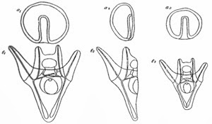
 a1 and b1: Normal gastrula and normal pluteus. a2 and b2: “Half”—gastrula and “half”—pluteus, that ought to result from one of the two first blastomeres, when isolated, according to the theory of “evolutio”. a3 and b3: The small but whole gastrula and pluteus, that actually do result. Driesch's drawing taken from his Gilfford Lecture.


His experiments contradicted the mosaic theory but also resulted in him abandoning science. Driesch renounced the study of developmental physiology and became a philosophy professor few years later, in 1909, proclaiming vitalism (the doctrine that living things cannot be explained by physical forces alone) until his death in 1941.
In 1897, Hans Spemann started experimenting with salamander embryos at the University of Würzburg in Germany. He had been inspired by Weismann's book The Germ Plasm: A Theory of Heredity which he had read one year earlier while guaranteed in the Alpes trying to recover from Tuberculosis. In 1902, trying to give an answer to the Roux-Driesch dispute he used a revolutionary, but yet rather simple method, to divide embryo cells by constructing a nose with his baby daughter's hair. Spemann tied this loop around the middle of the jelly-like membrane that salamander embryos have and squeezed it so that the two divided cells were formed into opposite halves of the egg's membrane. From these two newly formed cells two identical salamander embryos were developed both of them whole and normal. 
 Spemann had documented the first in-vitro animal clone! 
He continued working with salamander embryos and with Hilde Mangold they published a series of ingenuous experiments in 1924. In one of them they attempted the transfer of the nucleus of a sixteen-cell embryo to a single salamander embryo cell with no nucleus. The cell took up the nucleus and developed into a normal salamander. With this process, Spemann and Mangold completed one of the first cloning experiments using the nuclear transfer method.

Original pictures from Spemann's experiments. Top: drawing from the brain of a double headed embryo, 1902. Bottom: Double headed embryos after an incomplete constriction along the first cleavage turrow, 1900.


Unfortunately, Hilde Mangold died at the same year from a gas heater explosion in her house. She didn't live to see her thesis on the salamander embryos' work get published neither the Nobel Prize that was awarded to her mentor Hans Spemann in 1935 for their revolutionary work.


In 1938 Spemann published a book with his results entitled  Embryonic Development and Induction. He wondered whether cells that have already been differentiated would be able to form new organisms and speculated about the possibilty of creating a new organism by transferring the nucleus from a differentiated cell into an enucleated egg.  As he explained it: "Decisive information about this question may perhaps be afforded by an experiment which appears, at first sight, to be somewhat fantastical. This experiment might possibly show that even nuclei of differentiated cells can initiate normal development in the egg protoplasms."


......to be continued....

Further reading..
1. August Weismann: The Germ-Plasm: A Theory of Heredity translated by W. N. Parker in 1893
2.Dreisch's Gillford Lecture 
3. Dr. Ann A. Kiessling and Scott C. Anderson: Human Embryonic Stem Cells: An Introduction to the Science and Therapeutic Potential
4. Oscar Hertwig: The Biological Problem of Today